The familiar sound of clicking magnets returned to the CSU James L. Voss Veterinary Teaching Hospital on April 11, when the first patients were imaged using the newly installed, state-of-the-art Siemens Altea 1.5 Tesla Magnetic Resonance Imaging (MRI) machine.
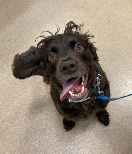
Griz, a spunky spaniel experiencing episodes of neck pain, was the first patient to be scanned. An MRI of the cervical spine (neck) is the gold standard diagnostic tool to provide the most information.
“MRI is essential to diagnose the cause of Griz’s clinical signs,” said Dr. Camilla Cooper, associate professor of neurology.
“Our physical examinations can tell us broadly where the neurological system is affected. However, they do not tell us what is causing the problem. Because it is so well protected, we can only look at most of the neurological system (most notably the brain and the spinal cord) by using advanced imaging.”
Patient No. 2
German shepherd Raylan was the second MRI patient of the day. Raylan’s family brought him to CSU after noticing he was dragging his right hind leg. A neurological exam indicated he likely had a lesion affecting the spinal cord somewhere between the shoulders and the hips. With his age, breed, neurologic exam, and history of chronic, slowly progressive symptoms, the neurology team’s primary concern was intervertebral disk herniation. MRI provided the best way to confirm exam findings and rule out less likely causes.
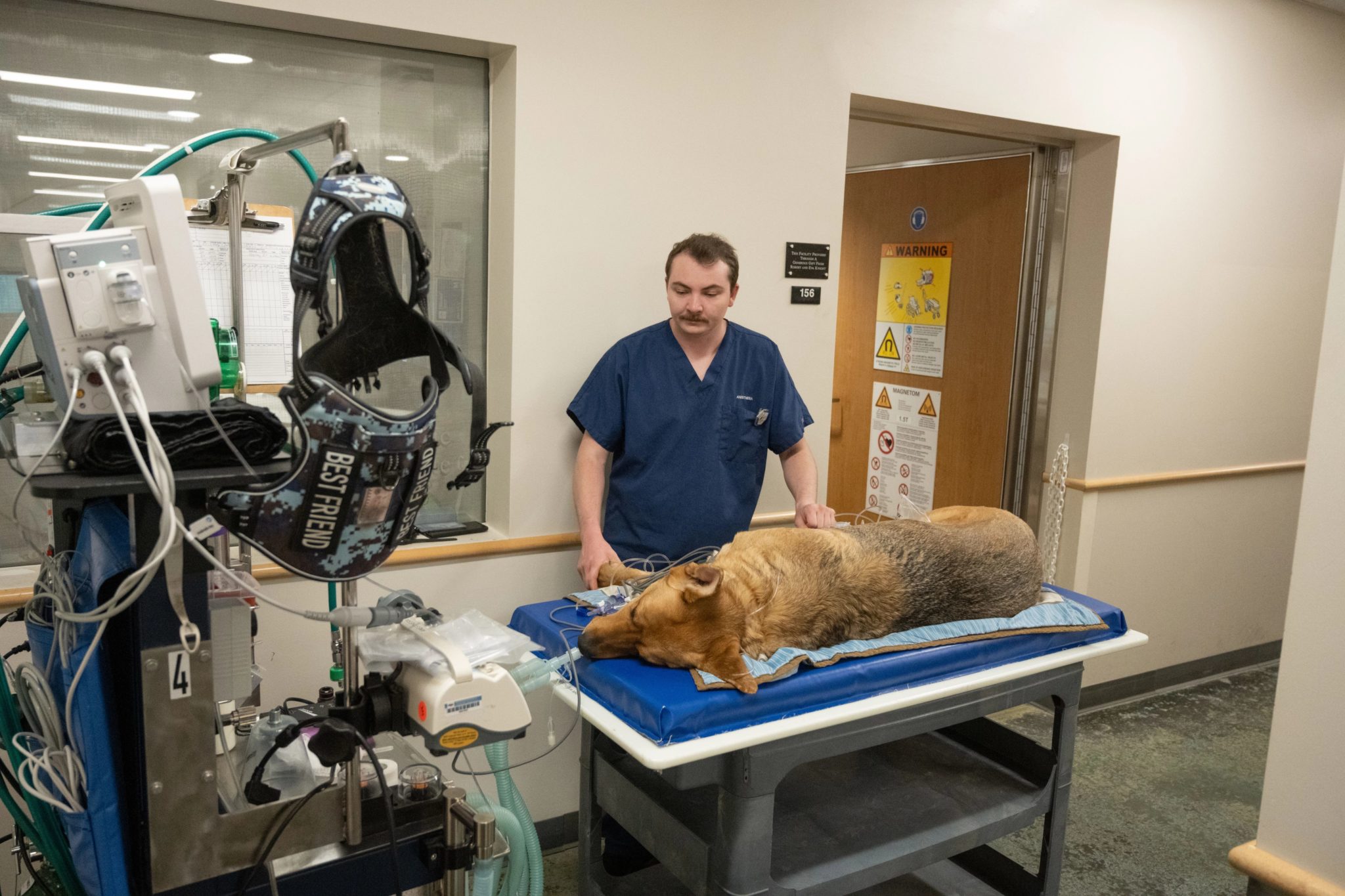
Raylan’s MRI revealed multiple degenerated disks in the spinal cord, confirming the initial intervertebral disk herniation diagnosis. Intervertebral discs are shock-absorbing anatomic structures between each vertebra in the spinal column. When these disks protrude, they can compress the spinal cord and cause neurologic abnormalities such as the ones exhibited by Raylan. Treatment options for Raylan include surgery or continued medical management for discomfort, exercise modification, and physical therapy. Presented with information and choices, Raylan’s family opted for medical management.
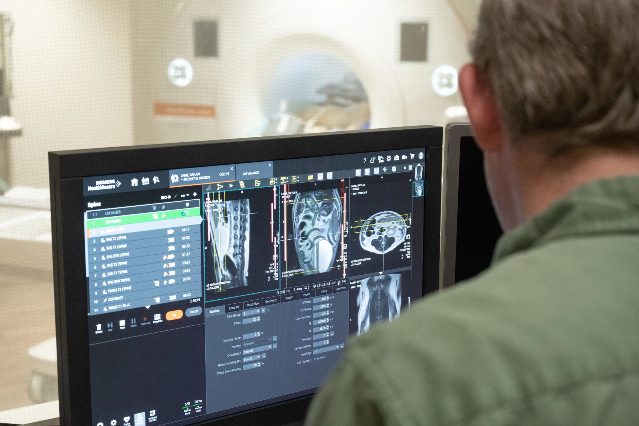
“The previous MRI was 20 years old, and although it gave great images, the technological advances mean that the new images are much better in detail. We can also run different types of sequences, which allow us to gain more information about any conditions we may find,” said Cooper.
New technology, new benefits for patients
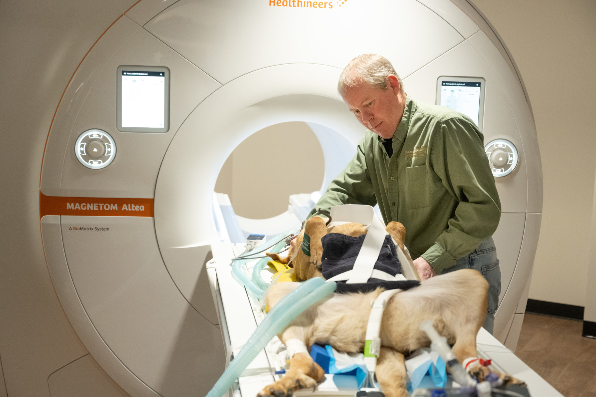
In addition to improved image quality and sequencing capabilities, the new MRI offers various additional benefits for patients. Positioning options are greatly enhanced for patient comfort, and the MRI’s larger bore (opening) allows the scanning of larger patients. And, with improved technology comes speed and efficiency, which means patients have shorter times under anesthesia.
“Another huge benefit to this scanner is that it acquires images much faster,” said Nikki Denard, advanced imaging lead for the CSU Veterinary Health System. “Faster scans are better for the patients, as it will shorten their anesthesia time and could help us see more patients in a day.”
According to Dr. Elissa Randall, professor of radiology, the imaging speed will take some getting used to. “Before I have a chance to read the scans, our technicians are asking if we want additional sequences or if the patient can leave the MRI suite. I used to have time to consult with our residents, read other images, or answer questions from staff. It’s a good problem to have!”
Neurology patients aren’t the only ones benefiting from advanced imaging capabilities. The new MRI includes a cardiology package, which may assist in diagnostics and surgical planning. Flexible coils that can wrap around almost any body part to help create an image also will expand musculoskeletal scanning capabilities.
“I am very excited about the future of imaging here at CSU, and I am glad to be a part of it,” said Stewart.