How does a thought occur? How is a memory stored? Do our memories require energy? These processes happen when neurons communicate by activating different synapses in the brain, and this communication requires energy.
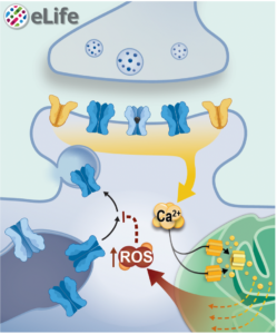
A new study from the lab of Fred Hoerndli, associate professor in the Department of Biomedical Sciences, looks at how neurons regulate and are regulated by synaptic mitochondria. They found that mitochondria can regulate the function of synapses, where brain cells communicate, via production of highly reactive chemicals, called reactive oxygen species, by mitochondria in response to the calcium that occurs when synapses are activated. Their results are published in eLife.
“This is the first study that clearly associates how the activity of the neuron can tailor reactive oxygen species, and that has an impact on how that neuron talks to other neurons at a very basic level,” said Hoerndli. “It was one of the coolest moments of my career when first author Rachel Doser showed me that she could excite a single mitochondria. Mitochondria are one of the building blocks of life, and we are finally asking: How do they talk?”
The lab previously found that reactive oxygen species can modify how glutamate receptors, prominent signaling molecules in the brain, get to synapses by acting on calcium regulation in the cell. They began this study wanting to know how reactive oxygen species may be involved in learning and memory on a molecular level and if mitochondria were involved in the process. This led the team to combine and optimize microscopy techniques to visualize glutamate receptors and mitochondria in combination with reactive oxygen species or calcium in live C. elegans worms.
They found that novel, activity-dependent increase of reactive oxygen species production by mitochondria decreased the amount of glutamate receptors arriving at the synaptic membrane. This calcium-dependent mitochondrial signaling may be protective against synaptic over-excitation and important for preventing excitotoxicity. The lab plans to continue studying this signaling in more detail to identify the key proteins involved. They also hope to address how the function, shape, and location of single synoptic mitochondria impacts neuronal communication using computational modeling.
“Mitochondria are well known as the powerhouse of the cell, but we know relatively little about their functions aside from the production of adenosine phosphate (ATP), the main energy-providing molecule in cells,” said first author and biomedical sciences PhD graduate Rachel Doser, now a postdoctoral researcher with CSU’s Department of Health and Exercise Science. “In this study, we hit on the leading edge of a newer idea that mitochondria are also signaling hubs. Uncovering these other functions of mitochondria, normally and in aging, is the new frontier.”