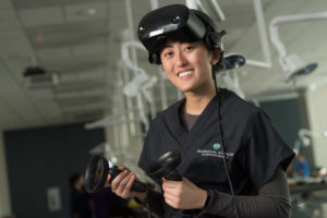
Goggles on, I stuck my head inside a patient’s chest and looked around as his lungs moved rhythmically with each breath. The image, made up of 10 CT scans taken in succession, was on display in three dimensions, plus the fourth dimension of time. If I were this patient’s surgeon, one of the infinite things I could do is measure the exact distance his lungs move when he breathes to find the best place to operate.
“For the first time ever, we can pull up any research or medical image, such as a CT or MRI scan, and in less than a minute be walking around inside of it,” said Tod Clapp, an assistant professor in the Department of Biomedical Sciences and head of its human anatomy program.
His team’s groundbreaking Human Virtual Anatomy Project, developed in 2017, is on a mission to revolutionize medical education. Along the way, it’s become apparent that it can do far more than that, and the team comes up with new ideas every day.
The project began with a desire to make the study of human anatomy more accessible, intuitive, and impactful for students by visualizing and manipulating magnified models of the brain, nervous system, and other structures of the body in all dimensions. It can now interact in depth with virtually any type of research or medical image, allowing specific parts to be manipulated, dissected from any plane, or viewed separately on command, such as by stripping away layers of skin, muscle, and bone to walk through heart valves or follow the path of brain signals. And one of the program’s most powerful tools is its capacity to have multiple people interact in the same virtual space at the same time, providing endless opportunities for students, teachers, clinicians, and researchers across the world to collaborate, teach, and learn.
The living teach the living
“When we talk about the program, people always ask, ‘Can you put it on a screen?’” Clapp said. “The answer is yes, but that would eliminate what virtual reality is. Seeing structure in two dimensions is never as informative as seeing it in three. We cannot fully explain the impact until they put the goggles on.”
And once they put the goggles on, it’s hard to get them to take them off. Three- and four-dimensional visualization and the ability to add and remove layers is not only very informative, it’s also mesmerizing. Clinicians report they’ve never seen anything that compares to virtual reality anatomy.“It gives them more perspective,” said Chad Eitel, the program’s lead developer. “Many clinicians find it helpful to look at things they don’t come across very often in their practice, or to look at really complicated things that are hard to conceptualize in three dimensions when you’re looking at a two-dimensional image.”
As part of a partnership with the Japan-based JSR Corporation, the team recently provided virtual reality setups to a variety of medical professionals who will explore using the program for pre-surgical planning, medical education, collaborating with other clinicians from a distance, and patient education, a novel and exciting potential direction that could offer patients the chance to become more informed and involved in their own health care than ever before.
“Countless colleagues have come in and spent hours exploring their own medical scans,” Clapp said. “It’s very exciting to them to walk through their own body. There is significant value in the patient education area because images in three dimensions are easier to understand. There’s no need to look at something in two dimensions when we can see it as it exists.”
The team is also collaborating with partners at the University of Florida who are using it for medical education, and with UCHealth, where clinicians are evaluating how it might provide useful insight that will help them do their jobs. Dr. Julie Dunn, a trauma and general surgeon at UCHealth, hopes to use it for better preoperative planning, which could save time and improve patient safety. “If this program improves both our ability to plan and stage various surgeries, it would be quite impactful,” Dunn said.
Aside from the obvious benefits to human and animal medicine, experts from a variety of professions are bringing data to the virtual anatomy team in order to explore how to view, play with, and study all kinds of things in an entirely new way, revealing that the potential applications of this program are truly limitless.
New health education outreach center goes virtual
Students started taking classes in the brand-new Health Education Outreach Center this spring. In a few months, the HEOC will house a first-of-its-kind virtual reality lab. Students are already using part of the virtual anatomy program on iPads, which allows them to interact with and dissect a three-dimensional human model and enhances the knowledge gained from their work with actual cadavers. By next fall, the team hopes to have the entire virtual reality program up and running in the new building and seamlessly integrated into the already robust curriculum, where it will drastically improve student learning and provide a world-class space for continuing medical education.
The Health Education Outreach Center provides needed space for greater numbers of students to pursue science and medicine; attracts new cohorts of future health care professionals to Colorado; and provides greater opportunities for public engagement. The center’s partnership with the National Western Center will bring its renowned hands-on educational outreach programs in health care and the life sciences to visiting students and families from around the world. The virtual reality lab will allow the new building to host a variety of continuing medical education programs and has already attracted interest from fields ranging from emergency medicine and nursing to dentistry and yoga teacher training.
“This is the benchmark facility for gross anatomy and neuroanatomy education in the nation,” said Tod Clapp, an assistant professor in the Department of Biomedical Sciences and head of its human anatomy program. “The new Human Virtual Anatomy Project will complement our robust curriculum. When students learn anatomy, they are learning about the importance of the relationships between structures, and you can’t truly do that anywhere but in a three-dimensional environment.”